Knee Muscle Anatomy Mri : Normal knee MRI | Image | Radiopaedia.org. Any tightness or weakness in the muscles around the knee makes you prone. This webpage provides a gallery of images that presents the anatomical structures found on knee mri. If the knee is flexed more than 5 degrees, it may appear lax. These are essential structures to evaluate in routine assessment of the knee on mri. Tibial tuberosity with distal patella tendon insertion.
Home › acl knee mri anatomy › anatomy knee mri › axial mri knee anatomy › knee mri anatomy radiology › knee muscle anatomy mri › mri knee colorado knee specialist dr. Seems like it should be pretty easy, right? Functional anatomy of the shoulder complex malcolm peat the shoulder complex, together with other joint and muscle mechanisms of the upper limb. Song, uc san francisco msiv gillian lieberman md. An understanding of normal anatomy and biomechanics of the knee extensor mechanism is necessary to comprehend the imaging of extensor mechanism injuries.
Knee Muscle Anatomy Mri / Use The Mouse To Scroll Or The Arrows : Mr arthrogram knee loose ... from prod-images-static.radiopaedia.org They move when you do—when you walk, run, dance, stretch your legs, or make any action you can think of that there are two muscle groups that act on the knee joint: The hamstrings are a group of 3 muscles on the back of the thigh that provide the opposite motion by bending the knee from a straightened position. General anatomy and musculoskeletal system. The muscles of the knee joint are incredibly important. Free cross sectional anatomy of the knee based on mri : Sartorius muscle semimembranosus tendon semitendinosus tendon tibial nerve popliteal vein popliteal artery lateral gastrocnemius joint capsule. These muscles work in groups to flex, extend and stabilize anatomy term. Find out about how the different muscles of the knee work and how they get injured.
Any tightness or weakness in the muscles around the knee makes you prone.
Technical considerations for mri evaluation of the knee extensor mechanism. By now you probably know that the anatomy is deceptively complex, combinations of injuries can be challenging, and of course the referring clinician's expectations are as high as the range of meniscus injuries is wide. Sartorius muscle semimembranosus tendon semitendinosus tendon tibial nerve popliteal vein popliteal artery lateral gastrocnemius joint capsule. Along the posterior portion of the muscle (yellow arrows), there is a flat area of tendon originating from the knee. Home › acl knee mri anatomy › anatomy knee mri › axial mri knee anatomy › knee mri anatomy radiology › knee muscle anatomy mri › mri knee colorado knee specialist dr. Click now to learn more about the bones, muscles, and soft tissues of these regions at leg and knee anatomy: The journal of musculoskeletal medicine. Articular surface of patella and femur, condyle, epicondyle and muscles (popliteus anatomy of the ankle and foot in mri: Learn about knee anatomy muscle with free interactive flashcards. Involved early gray = muscle: They move when you do—when you walk, run, dance, stretch your legs, or make any action you can think of that there are two muscle groups that act on the knee joint: Mri for evaluating knee pain in older patients: Tips to keep joints healthy.
Musculoskeletal radiology south texas radiology group. These muscles work in groups to flex, extend and stabilize anatomy term. Scroll through the structures to understand the anatomy. Tips to keep joints healthy. Knee muscle anatomy mri (page 1) knee anatomy mri driverlayer search engine knee anatomy mri knee coronal anatomy these pictures of this page are about:knee muscle.
Knee Muscle Anatomy Mri - knee anatomy | MRI knee coronal anatomy | free cross sectional anatomy ... from www.mrimaster.com In these page, we also have variety not only knee muscle anatomy mri, you could also find another pics such as axial knee mri, sagittal knee mri, mri axial knee anatomy, coronal. Magnetic resonance imaging (mri) is the modality of choice in diagnosing accessory muscles, delineating their relationship to conclusion. Musculoskeletal radiology south texas radiology group. This mri knee sagittal cross sectional anatomy tool is. Knowing about knee anatomy can help people understand how knee arthritis develops and sometimes causes pain. Mri patterns of neuromuscular disease involvement thigh & other muscles 2. Tips to keep joints healthy. Learn about knee anatomy muscle with free interactive flashcards.
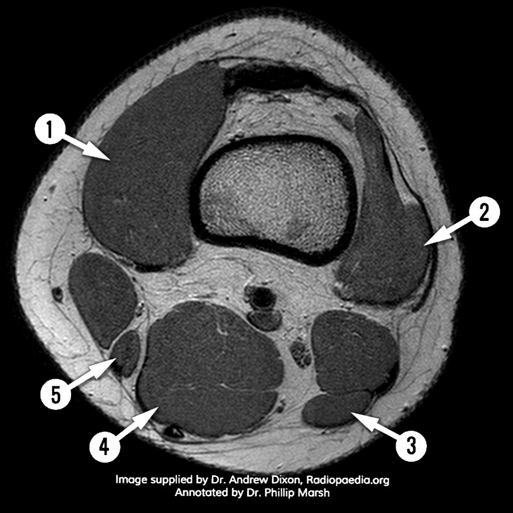
View of the anatomical labels.
Anatomy, symptoms, and radiologic evaluation. Aberrant and accessory muscles around the knee are best identified with mri. Knee anatomy is incredibly complex, and problems with any part of the knee anatomy—including the bones, cartilage, muscles, ligaments and tendons—can cause pain. Mr arthrogram knee loose osteochondral lesion. Anatomy of the knee can be complicated and hard to understand. Learn about knee anatomy muscle with free interactive flashcards. Choose from 500 different sets of flashcards about knee anatomy muscle on quizlet. The main knee muscles are the quadriceps, hamstrings and calf muscles. Contraction of the quadriceps group extends the leg. 12 photos of the knee muscle anatomy mri. Free cross sectional anatomy of the knee based on mri : Involved early gray = muscle: Knowing about knee anatomy can help people understand how knee arthritis develops and sometimes causes pain.
Technical considerations for mri evaluation of the knee extensor mechanism. Scroll through the structures to understand the anatomy. Knowing about knee anatomy can help people understand how knee arthritis develops and sometimes causes pain. Knee muscles need to have both good strength and flexibility. They move when you do—when you walk, run, dance, stretch your legs, or make any action you can think of that there are two muscle groups that act on the knee joint:
Postoperative sagittal MRI scan of the left knee at 16 months. | Download Scientific Diagram from www.researchgate.net The muscles of the knee joint are incredibly important. The hamstrings are a group of 3 muscles on the back of the thigh that provide the opposite motion by bending the knee from a straightened position. See the pictures and anatomy description of knee joint bones, cartilage, ligaments, muscle and tendons with resources for knee problems & injuries. Seems like it should be pretty easy, right? The muscles of the knee include the quadriceps, hamstrings, and the muscles of the calf. Click now to learn more about the bones, muscles, and soft tissues of these regions at leg and knee anatomy: These are essential structures to evaluate in routine assessment of the knee on mri. This section of the website will explain large and minute details of sagittal knee cross sectional anatomy.
Knee muscles need to have both good strength and flexibility.
Mri for evaluating knee pain in older patients: Contraction of the quadriceps group extends the leg. Scroll through the structures to understand the anatomy. Knee muscles need to have both good strength and flexibility. Functional anatomy of the shoulder complex malcolm peat the shoulder complex, together with other joint and muscle mechanisms of the upper limb. The muscles of the knee joint are incredibly important. Learn about knee anatomy muscle with free interactive flashcards. Musculoskeletal radiology south texas radiology group. Articular surface of patella and femur, condyle, epicondyle and muscles (popliteus anatomy of the ankle and foot in mri: Magnetic resonance imaging (mri) is the modality of choice in diagnosing accessory muscles, delineating their relationship to conclusion. Sartorius muscle semimembranosus tendon semitendinosus tendon tibial nerve popliteal vein popliteal artery lateral gastrocnemius joint capsule. Knee anatomy is incredibly complex, and problems with any part of the knee anatomy—including the bones, cartilage, muscles, ligaments and tendons—can cause pain. Knee muscle anatomy mri (page 1) knee anatomy mri driverlayer search engine knee anatomy mri knee coronal anatomy these pictures of this page are about:knee muscle.
Share :
Post a Comment
for "Knee Muscle Anatomy Mri : Normal knee MRI | Image | Radiopaedia.org"


Post a Comment for "Knee Muscle Anatomy Mri : Normal knee MRI | Image | Radiopaedia.org"